
3D/4D sonography uses ultrasound waves to create three-dimensional still images (3D) and real-time moving images (4D) of the fetus. It helps visualize fetal anatomy, movements, and development more clearly than standard 2D scans.
Detailed anatomical assessment during pregnancy
Better visualization of fetal face, limbs, and movement
Helps in counseling parents and assessing suspected anomalies
Expectant mothers with routine prenatal care needs, those with prior inconclusive scans, or when the doctor requests detailed anatomical views.
No special preparation for most 3D/4D scans.
For early pregnancy ultrasounds, a full bladder may be requested — follow appointment instructions.
Wear comfortable clothing and bring previous ultrasound reports if available.
Non-invasive, painless, and uses no ionizing radiation.
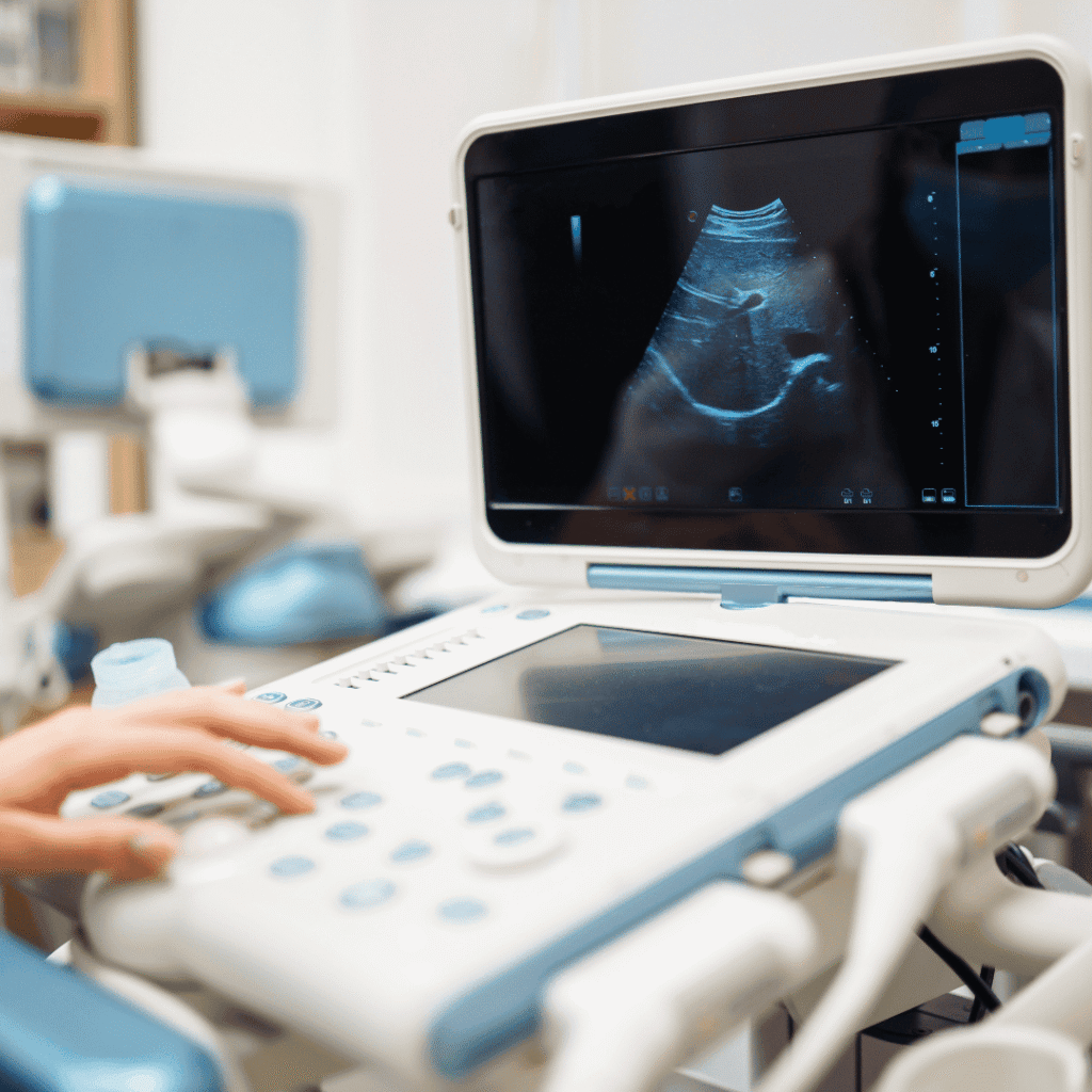
You’ll lie on the exam bed while a transducer is moved on the abdomen.
The session lasts ~20–45 minutes depending on views required.
Preliminary findings discussed in person.
Detailed imaging/report available within 24–48 hours. Digital copies can be sent via WhatsApp/email.
Experienced sonographers, modern ultrasound machines, and a calm, private environment for your scan
Is 3D/4D ultrasound safe? — Yes, it uses sound waves and is considered safe at recommended exposure levels.
When in pregnancy is it best? — Often done between 20–32 weeks. Your doctor will advise the best time.
Can family view the scan? — Yes — we welcome a companion. Please check hospital guidelines during your visit.
Trusted diagnostic centre in Vikhroli offering accurate imaging, pathology tests, and expert sonography services.